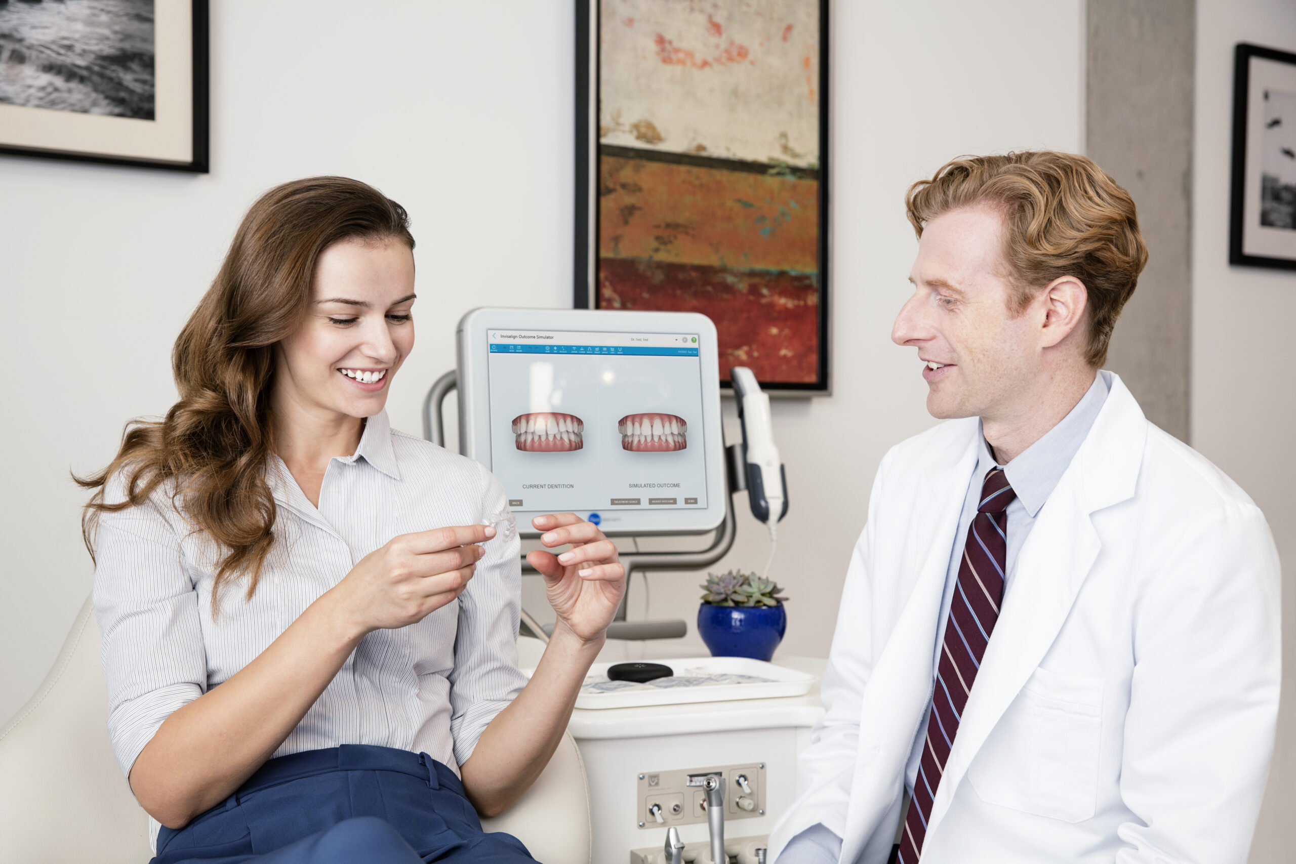
Thermal imaging is rapidly becoming an essential tool in modern dentistry. It provides a non-invasive and highly effective way to detect oral conditions that may otherwise go unnoticed during traditional examinations. By using thermal imaging to assess temperature variations in oral tissues, dentists can gain valuable insights into a patient’s oral health. This cutting-edge technology helps with early detection of inflammation, infection, and other dental conditions, significantly improving treatment outcomes and patient care.
In this article, we will explore the benefits of thermal imaging in dentistry, its use as a diagnostic tool, and how it can help detect inflammation and other issues at the earliest stages.
What is Thermal Imaging in Dentistry?
Thermal imaging, also known as infrared thermography, is a diagnostic technique that uses specialized cameras to detect and record temperature variations on the surface of the body. In dentistry, thermal imaging cameras create heat maps that provide visual representations of temperature differences in the tissues of the mouth. This technology can identify areas of increased heat or cooler regions, which may be indicative of underlying oral health problems.
The main advantage of thermal imaging is its ability to detect changes in temperature that are associated with various oral conditions, often before they become visible to the naked eye or are detectable through traditional diagnostic methods.
Benefits of Thermal Imaging in Dentistry
1. Non-Invasive and Painless
One of the primary benefits of thermal imaging is that it is completely non-invasive and painless for patients. Traditional diagnostic methods like X-rays or probing can sometimes cause discomfort or require the use of contrast agents or other invasive procedures. Thermal imaging eliminates this discomfort by simply scanning the surface of the mouth without any physical contact or need for injections, making it an ideal choice for patients of all ages.
2. Early Detection of Inflammation and Infection
Thermal imaging can reveal subtle temperature changes that are often the first signs of inflammation or infection in the mouth. Conditions such as periodontal disease, abscesses, or infections in the teeth and gums often present with increased heat in the affected areas due to increased blood flow and immune response. By detecting these changes early, thermal imaging allows dentists to diagnose conditions before they progress, enabling faster and more effective treatments.
For example, periodontitis, a common form of gum disease, can be identified early through thermal scans that highlight areas of infection that may not yet show visible symptoms.
3. Enhanced Diagnostic Accuracy
Thermal imaging can increase the accuracy of oral diagnostics by providing an additional layer of information. It complements traditional diagnostic tools, such as X-rays, allowing the dentist to confirm or refine their diagnosis. While X-rays reveal structural changes, thermal imaging highlights temperature patterns that are directly linked to inflammation and tissue damage. This helps in making more informed decisions about treatment and care.
4. Early Detection of Cavities and Tooth Decay
Dental caries (cavities) are typically detected through physical exams and radiographs, but thermal imaging provides a different and valuable perspective. Decay often leads to changes in the tooth’s temperature due to increased bacterial activity and fluid accumulation. Thermal imaging can detect these temperature changes in the tooth’s surface, allowing dentists to identify early-stage cavities that might not yet show up on X-rays or during visual inspections.
5. Assessing Bone Health Around Implants
Thermal imaging is also useful in implant dentistry. When dental implants are placed, the surrounding bone should be healthy and stable to ensure long-term success. Thermal imaging can detect areas of poor circulation or infection around the implant site that may indicate bone loss or complications. Early detection of these issues can prevent implant failure and allow for timely interventions.
6. Monitoring Treatment Progress
Thermal imaging can also be used to monitor the progress of treatments for conditions like root canal infections, gum disease, and post-surgical recovery. By comparing thermal images taken at different stages of treatment, dentists can assess how well a condition is responding to therapy and make adjustments if necessary. This helps optimize treatment plans and ensures better patient outcomes.
7. Improving Patient Comfort and Trust
Many patients feel anxious about dental visits, especially when they involve potentially uncomfortable procedures like X-rays or deep cleanings. Since thermal imaging is non-invasive and involves no discomfort, it can help alleviate patient fears and build trust. Additionally, by providing immediate results, patients can see the value of the technology firsthand, which can increase their confidence in the diagnosis and treatment plan.
How Thermal Imaging Works in Dentistry
Thermal imaging in dentistry uses infrared cameras that detect infrared radiation (heat) emitted from the surface of the body. These cameras create a visual representation, or thermal image, by translating the temperature variations into a color gradient. Warmer areas appear as lighter colors (often red or yellow), while cooler areas are displayed in darker colors (like blue or purple).
Steps in Using Thermal Imaging for Diagnosis:
Initial Examination: The dentist conducts a routine clinical exam and identifies any areas of concern, such as pain, swelling, or sensitivity.
Thermal Scan: A thermal imaging camera is then used to scan the areas of interest, such as the teeth, gums, or jaw. The camera detects subtle changes in temperature that may indicate infection, inflammation, or tissue damage.
Analysis: The dentist analyzes the thermal images, looking for areas of increased heat or temperature differences. These areas are then correlated with potential diagnoses, such as infection, abscesses, or gum disease.
Further Action: Based on the findings, the dentist may decide to follow up with traditional diagnostic tools (like X-rays or biopsies) or proceed with treatment if a clear condition is identified.
Applications of Thermal Imaging in Dentistry
Detection of Periodontal Disease: Identifying early signs of gum disease and inflammation.
Cavity Detection: Revealing temperature changes associated with tooth decay.
Root Canal Diagnosis: Detecting infection or inflammation in the tooth’s root.
Implant Monitoring: Assessing the health of bone and tissue surrounding dental implants.
Post-Surgical Care: Monitoring healing and recovery after oral surgeries.
Thermal imaging is a revolutionary tool in dentistry that offers a non-invasive, accurate, and efficient way to diagnose and monitor various oral conditions. From detecting inflammation and infection to helping with early cavity detection and post-surgical monitoring, the benefits of thermal imaging are immense. As dental technology continues to evolve, thermal imaging is expected to play an even larger role in ensuring patients receive the best care possible, improving diagnostic accuracy, and enhancing overall treatment outcomes.
By incorporating thermal imaging into daily practice, dentists can provide more precise, efficient, and comfortable care for their patients, contributing to better oral health and more successful dental treatments.

